 Pixolite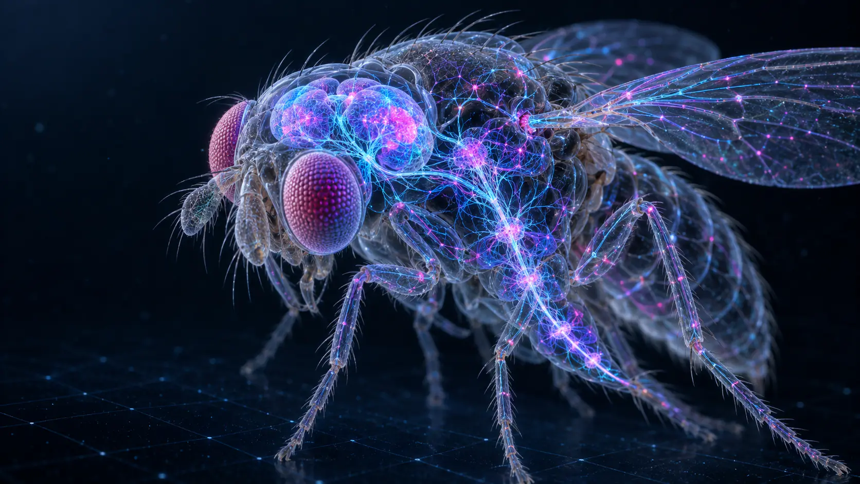
A fruit fly’s brain is smaller than a poppy seed. Yet inside it sits a network capable of seeing, navigating, remembering, choosing food, escaping danger and performing surprisingly elaborate courtship rituals.
Scientists can now inspect that network neuron by neuron.
On 3 September 2026, Google Research announced the first complete wiring diagram, or connectome, of an adult male fruit fly’s brain and central nervous system. Created with the Howard Hughes Medical Institute’s Janelia Research Campus and an international group of researchers, the map contains precisely 166,691 neurons, approximately 125 million synaptic connections and 11,691 identified cell types.
It includes the optic lobes, central brain and ventral nerve cord, the insect equivalent of a spinal cord. By number of neurons, Google calls it the largest complete brain map produced so far. The Google Research announcement and the paper published in Cell provide the full scientific details.
The achievement has serious scientific value. It has also produced one of the more entertaining technology stories of the year: within days, independent developers were connecting versions of the fly network to Doom, Super Mario 64, Minecraft, Beat Saber and cryptocurrency markets.
That combination of rigorous neuroscience and gleeful experimentation shows why an open brain map can be so powerful. It also makes it important to understand what has actually been mapped.
How do you map a brain this small?
Researchers first imaged the nervous system using seven enhanced focused-ion-beam scanning electron microscopes. Over 13 months, the instruments generated a 160-teravoxel image at a resolution of eight nanometres.
Google’s machine-learning systems then helped turn the enormous collection of images into three-dimensional neurons. The main technique involved flood-filling networks: neural networks that begin at one point in an image and identify the neighbouring pixels belonging to the same biological structure.
This automation was essential, but it was not the end of the process. Human specialists at Janelia proofread the reconstructed neurons, corrected errors, classified cell types and checked connections. The result is better understood as an extraordinarily detailed anatomical atlas than as a brain captured in software.
The complete dataset is openly available under a Creative Commons licence. Researchers can explore it visually, search connections through neuPrint or download the underlying synapses, annotations and neuron skeletons for their own analysis. Janelia’s MaleCNS portal provides these tools and downloads.
Comparing male and female brains
A complete adult female fruit-fly brain was published through the FlyWire project in 2024. That map contains 139,255 proofread neurons and more than 50 million synapses. Scientists can now compare the two sexes at single-synapse resolution for the first time. FlyWire documents the earlier map and the growing body of research built upon it.
Most of the male and female nervous systems are remarkably similar. The new study matched more than 96% of neurons in the male brain to cell types found in female datasets. The researchers nevertheless identified 262 male-specific, 69 female-specific and 114 sexually dimorphic cell types: cells that exist in both sexes but differ in number, shape or connectivity.
These differences are concentrated in higher brain centres rather than basic sensory and motor regions. Male and female flies largely use similar equipment to see and move, while some of the circuits that interpret information and select behaviour are wired differently.
That finding gives researchers a powerful route into questions about courtship, mate selection, aggression and sex-specific movement. The map reveals circuit switches capable of routing similar sensory signals towards opposing behaviours. Although visibly sex-specific neurons form only a small portion of the brain, their connections can influence much larger networks. Janelia’s research summary explains how these differences affect about 4.8% of the male central brain.
Turning anatomy into testable biology
A connectome allows researchers to trace a possible signal from a sensory receptor through intermediate neurons and into cells controlling muscles. It makes questions that once seemed impossibly broad much easier to investigate:
- How does the smell or sight of food produce movement?
- How does a fly choose between approaching sugar and avoiding something bitter?
- Which circuits make it groom a dirty antenna?
- How does it steer, take off, land or escape a looming object?
- How do hunger, sleep, mating status or previous experience alter those decisions?
Earlier work using the female connectome has already demonstrated the method. In one study, scientists constructed a simplified computational model of the whole brain and stimulated virtual taste or touch neurons. The model correctly predicted cells involved in feeding and antennal grooming. Researchers then activated some of those cells in living flies and confirmed the predicted behaviour.
The peer-reviewed Nature study showed that a structural map, combined with neurotransmitter information and simple neural dynamics, can generate hypotheses that work in real animals.
Another team built a network constrained by the fly’s visual wiring. After training it to estimate motion, the model predicted how individual types of visual neuron respond to moving scenes. This suggests that connectomes can help scientists work backwards from anatomy to computation: not merely asking where neurons connect, but discovering what those connections calculate. That research also appeared in Nature.
Fruit-fly connectomes are now being used to study vision, smell, taste, navigation, feeding, grooming, sleep, circadian rhythms, pain, learning, ageing and escape behaviour. Because flies can be genetically altered with exceptional precision, researchers can use the map to identify a circuit and then switch selected neurons on or off in a living animal.
The work may eventually inform human neuroscience, but the route is indirect. Fruit flies share many fundamental genes, neurotransmitters and organisational principles with larger animals. Discovering how a complete nervous system fails during ageing or disease can suggest mechanisms to test in mammals. It does not make the fly connectome an immediate blueprint for treating a human brain.
From biological wiring to robots
Computer scientists are also treating connectomes as possible designs for artificial intelligence.
One recent experimental system, FlyGM, converts a fly connectome into a message-passing network attached to a biomechanical fly body. Its researchers report that the resulting controller can learn walking, turning and flight tasks more efficiently than comparison networks with randomised wiring.
Another project, FLYNN, uses fruit-fly brain topology in a navigation network and reports better resilience when a robot loses visual information. Both are early research projects rather than established commercial systems, but they test an intriguing proposition: evolution may have discovered compact, robust network structures that engineers can reuse. The FlyGM and FLYNN papers describe the experiments.
The connectome has also been implemented on Intel’s Loihi 2 neuromorphic hardware, whose electronic architecture is designed to resemble networks of spiking neurons. Such experiments could contribute to smaller, more energy-efficient control systems for robots and autonomous devices.
Why is the fly playing Doom?
The stranger applications began appearing almost immediately after Google publicised the map.
In DOOMFLY, game frames are converted into artificial signals for thousands of visual neurons. Activity travels through a simplified model built from the MaleCNS wiring, and selected output neurons are translated into commands such as move, turn and fire. Damage produces an artificial aversive signal in dopamine cells.
It is a clever, open experiment, but its creator is unusually clear about the limitations: the sensory interface, neural dynamics, reinforcement mechanism and game controls are engineered approximations. The current system has not demonstrated that it learned to survive. It is therefore more accurate to say that a simulation shaped by fly anatomy is controlling Doom than that a fly’s mind understands the game. The DOOMFLY repository publishes its methods, assumptions and negative results.
The Super Mario 64 project follows a similar loop. Images from Mario’s environment are presented to modelled fly vision, activity runs through the network and selected outputs become controller actions. Its documentation explicitly describes it as an experiment using real wiring and simple rules, rather than a validated fly or trained Mario player. The project is available on GitHub.
Other developers have placed connectome-based controllers inside Minecraft creatures and Beat Saber demonstrations. Some are using the network to steer virtual flies, drones or simulated bodies. A few projects translate financial charts into visual or environmental stimuli and turn neural output into buy, sell or hold decisions.
These trading experiments have produced eye-catching headlines, but there is no evidence that fruit-fly wiring can predict markets. They are creative interfaces built around a biological graph, not investment systems.
A wiring diagram can constrain a simulation and inspire new experiments. On its own, it does not recreate the living animal whose connections were mapped.
A map is not a mind
A connectome records which neurons appear to connect and, in many cases, which neurotransmitter they probably use. It does not fully record the electrical state of every cell, the exact strength and timing of every synapse, changing chemical concentrations, hormones, gene activity, learning history or the continuous feedback between brain and body.
The original fly also had to be killed and physically sectioned for electron-microscope imaging. Nothing conscious or living was extracted and placed online.
The comparison is similar to having an extremely detailed road map without knowing the location, destination or behaviour of every vehicle. The roads constrain what can happen, and their design tells us a great deal about the system, but the map alone does not recreate the traffic.
That limitation does not diminish the achievement. It defines the next stage of the work: combining anatomy with recordings of neural activity, gene expression, body mechanics and observed behaviour. As those layers are added, researchers can build increasingly useful digital models for testing predictions before performing difficult experiments on living animals.
What comes after the fruit fly?
Google is already applying related mapping technology to portions of zebrafish, mouse and human brains. The scale rises sharply: Google estimates that a mouse brain is roughly a thousand times larger than the fly dataset, while a human brain is another thousand times larger. A complete human synaptic connectome therefore remains far beyond present capabilities. Google’s neural-mapping programme outlines those projects.
For now, the fruit fly occupies a valuable middle ground. Its nervous system is small enough to map, yet complicated enough to produce memory, navigation, flexible decisions and social behaviour. The new male connectome gives science its most detailed opportunity yet to follow those behaviours from sensory input, through a complete brain, and out towards the body, even if the internet’s first instinct was to send it into Doom.
Research and project status checked on 14 September 2026.
If you have an ambitious technology idea and need help turning it into a clear, reliable digital product, book a direct call with Damian. Pixolite designs and builds websites, applications and custom software around real business goals.